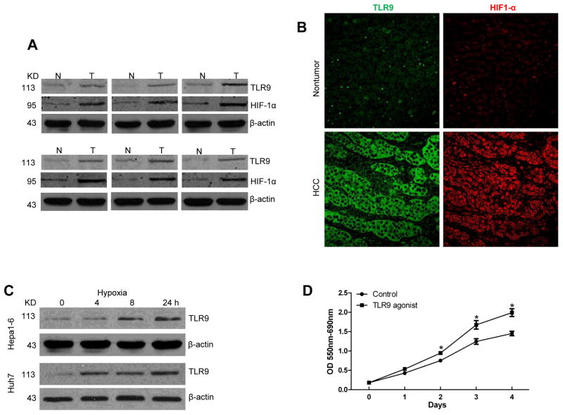
Fig. 1. TLR9 is overexpressed in hypoxic HCC and promotes tumor growth.
(A) TLR9 and HIF-1α protein tissue levels were measured using Western blot analysis in paired HCC samples and their non-tumor liver counterparts. (N, nontumor liver; T, tumor). Selected samples are representative of 24 unique paired samples. (B) Representative images of the nontumor and HCC tissues stained for TLR9 and HIF-1α (Green, TLR9; red, HIF-1α). (C) TLR9 protein expression in Hepa1-6 and Huh7 cells subjected to a time course of hypoxia (1% O2). (D) MTT assay at different time points of hep1-6 cells after TLR9 agonist treatment. Data is presented as mean±SE and is representative of 3 independent experiments. *p< 0.05.

