
Figure 3. Calcineurin inhibitor dephosphorylates cofilin during OGD.
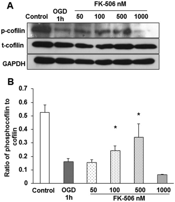
Differentiated PC12 neuronal cells were pretreated with different concentrations of calcineurin inhibitor (FK-506) for 1 h and then subjected to 1 h of OGD. A. The decreased levels of phosphocofilin in OGD were found to be restored with 100 and 500 nM concentrations of the inhibitor. B. The bar graph represents the cumulative result of three experiments; densitometric values of the bands were calculated using Image J software. The ratio of phosphocofilin to cofilin levels were plotted against the different treatment groups. The data was expressed as mean±SEM, where p<0.05 was considered significant. *vs 1 h of OGD.