
Figure 7. Cofilin dynamics during ischemia.
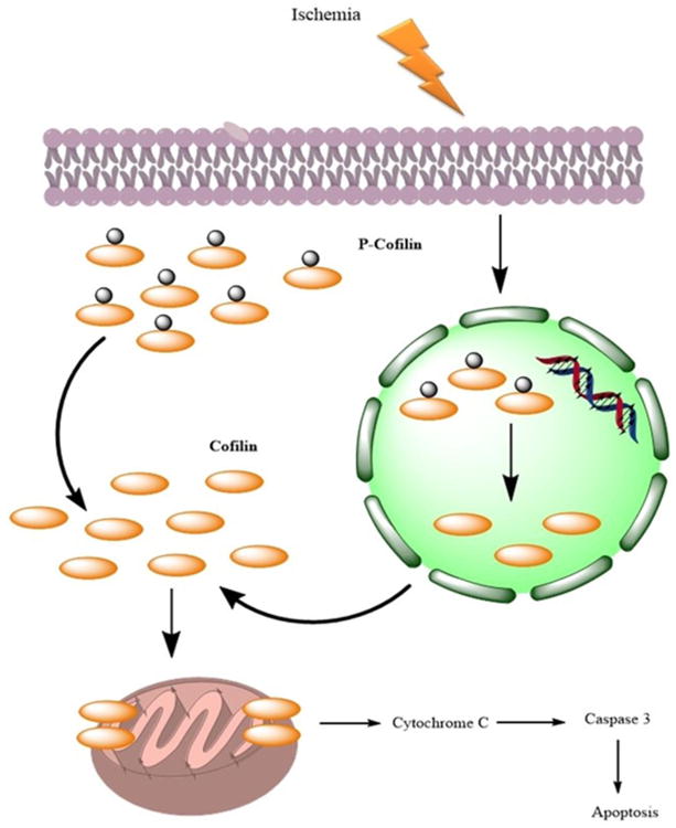
In normal conditions, a large proportion of cofilin exists in the nucleus and cytosol in phosphorylated form (phosphocofilin). However, during ischemia, nuclear phosphocofilin is dephosphorylated and translocated to the cytosol and then subsequently to the mitochondria, where it induces cytochrome C release and subsequent apoptosis.