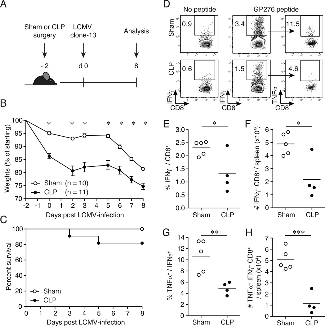
Figure 1.
Increased susceptibility to LCMV clone-13 infection and impaired primary Ag-specific CD8+ T cell responses in septic mice. (A) Experimental model. Two days prior to LCMV clone-13 infection (2 × 10⁶ PFU/mouse; i.v.), CLP or sham surgery was performed on inbred C57BL/6 (Thy1.2/1.2) mice. Subsequent CD8+ T cell analyses and viral titers were examined on day 8 post-LCMV clone-13 infection. (B) Morbidity and (C) survival rate were analyzed on indicated days post-CLP or sham surgery. (D) Representative flow cytometry plots illustrating expression of IFNγ+ of CD8+ T cells and TNFα+ of IFNγ+ CD8+ T cells. Numbers within plots represent the frequency of cytokine positive as indicated. Summary data of (E) frequency and (F) total number of IFNγ+ CD8+ T cells. Ex vivo GP276-peptide stimulation of splenocytes on day 8 post-LCMV clone-13 infection. Summary data of (G) frequency and (H) total number of TNFα+ IFNγ+ CD8+ T cells. Dots represent individual mice. Data analyzed in morbidity curve by two-tailed, unpaired Student t test (10–11 mice/group). Data analyzed in survival curve by Gehan-Breslow-Wilcoxon test, no statistical significance found. Ag-specific CD8+ T cell responses analyzed by two-tailed, unpaired Student t test (4–5 mice/group). Data are representative of three to four independent and similar experiments. * p < 0.05, ** p < 0.01, *** p < 0.0001.

