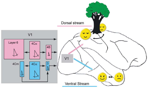
Figure 4.

Motion-selective pathways in V1 and their relationships to the dorsal and ventral cortical streams of information processing. The dorsal and ventral streams are indicated on a drawing of a lateral view of a monkey brain. Rectangular colored boxes represent median values of physiological characteristics of direction selective cells in individual V1 layers. Cells were considered direction selective if they fired twice as many spikes in the preferred as in the nonpreferred direction. Box width is proportional to measured receptive field width. Box height is proportional to the percentage of cells responsive to a bar longer than 60 min. The symbol for included angle is proportional to the measured half bandwidth of the orientation tuning curve. Layer 4Cα feeds layer 4B and layer 6 in the pathway leading to the dorsal stream (magenta). Layers 4B and 6 send outputs from V1 through area MT to the parietal areas (magenta arrow) responsible for sensing object motion and location, including relations in depth. Layer 4Cα also feeds layers 4Cm and layer 3 in a separate direct pathway to V2, which projects to the ventral stream (cyan) responsible for object recognition, including faces. The small dimensions of layer 3 receptive fields (narrow widths as well as preferences for very short stimuli) are well suited to sense subtle motions within objects such as changes in facial expressions. (From Gur and Snodderly, 2007).
