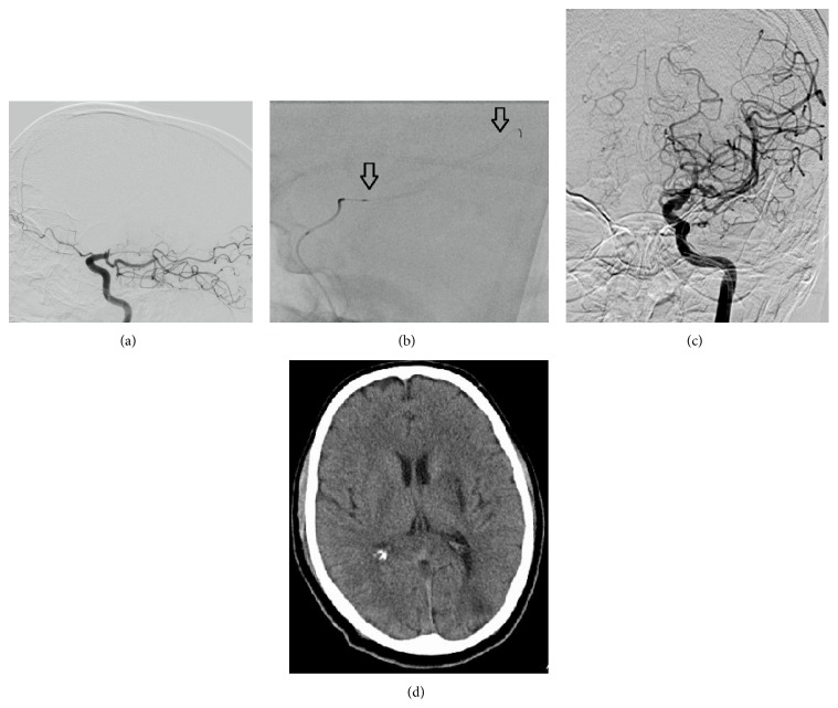
Figure 2.
(a) DSA reveals persistent carotid T occlusion and fresh thrombus on the occluded segment. Fetal PCA is observed on the lateral projection (black arrow). (b) Deployed Trevo-ProVue device (distal and proximal markers = black arrow). (c) After two passes with Trevo device, successful recanalization was achieved (TICI 2b). Both anterior cerebral arteries are filled from right hemisphere. (d) Control CT scan shows small infarction on basal ganglia.

