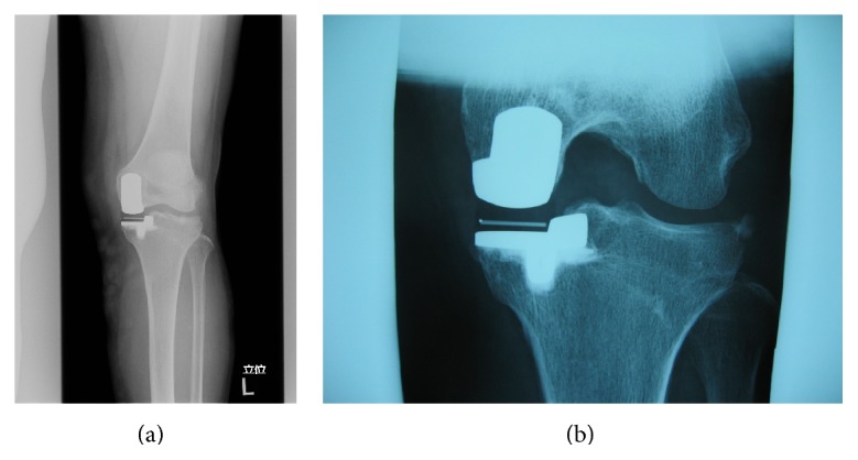
Figure 5.

Malrotation of femoral rotation (simulation on Athena). The simulated malrotation seemed to be satisfying on simulated anterolateral view (a), although the femoral component (contour) is set according to the shape of the medial condyle on axial view of CT scan (b). (This sample is neither of the cases.)
