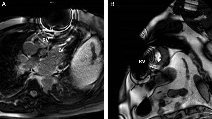
Figure 3.
Representative magnetic resonance imaging artefacts in patients with a left thoracic cardiovascular implanted electronic device. (A) Four-chamber view. (B) Short-axis view. Image distortion/void hampering diagnostic image quality particularly regarding the right ventricle and anterior wall of the left ventricle is apparent. RV, right ventricle; LV, left ventricle.

