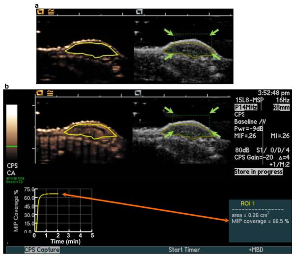
Fig. 1.
Setup for real-time maximum intensity persistence (MIP) ultrasound imaging with motion compensation. Motion compensation was accomplished by manually placing a tracking box (green dotted line; green arrows) over the subcutaneous human colon cancer xenografts located on the back of mice (a). A region of interest (ROI; yellow outline of tumor) was drawn on both contrast mode (left) and B-mode (right) images before contrast agent injection for real-time measurement of MIP percent contrast area. b MIP percent contrast area in the ROI was calculated in real-time after contrast reached equilibrium and was displayed graphically on the ultrasound screen (orange arrow). (Color figure online)

