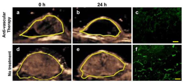
Fig. 4.

Representative two consecutive (24 h apart) transverse MIP ultrasound images of a human colon cancer xenograft in a mouse with (a, b) and in another mouse without (d, e) anti-vascular therapy using a vascular disruptive agent. Note substantial decrease in measured MIP percent contrast area in treated xenograft and no significant change of MIP percent contrast area in non-treated xenograft. Ex vivo microvessel density analysis confirmed decreased angiogenesis in treated (c) xenograft and no significant change in non-treated xenograft (f). Yellow scale in c and f = 100 μm. (Color figure online)
