
Fig. 3.
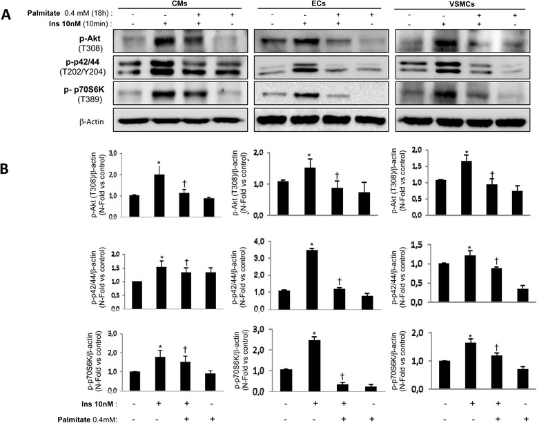
Palmitate induced cardiovascular insulin resistance. Western blot analysis (a) and its quantifications (b) of Akt phosphorylation (T308), p42/44 (T202/Y204) and p70S6K (T389) induced by insulin (10nM, 10 min) in presence or absence of oleate (2 h) in CMs, ECs and VSMCs. β-actin was used as charge control. *p < 0.05 vs. control; †p < 0.05 vs. stimulus