
Figure 5.
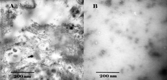
Electron micrographs of A: tau protein without crocin; B: tau protein with crocin. Samples were applied to carbon-coated gold grids, negatively stained with 2% uranyl acetate and analyzed by H600 transmission electron microscope (Hitachi Co.) operating at 50,000× at 75 kV excitation voltages. After incubation for 120 hr tau protein without crocin sample showed mature fibers as well as amorphous aggregates. In the presence of crocin, the majority of tau proteins was amorphous form. The Scale bar represents 200 nm