
Figure 10.
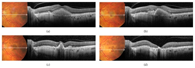
Serial color photos and OCT scans of a patient with patchy pattern during a 5-year follow-up. (a) At baseline examination, yellowish vitelliform-like material was visible on fundus photo; OCT scan showed that the material was homogeneous, mildly hyperreflective, and localized in the subretinal space above the RPE. It was associated with a diffusely thickened inner segment/outer segment junction and intact external limiting membrane. (b) After 1 year, the vitelliform material started resorbing inferonasally to the fovea; on OCT scan, the subretinal material was no more homogeneous. (c) At year 3, the vitelliform material shrunk, and initial RPE and photoreceptors atrophy were appreciated perifoveally. (d) At year 5, the vitelliform material was completely resorbed; OCT scan showed absence of external limiting membrane, marked thinning of the outer nuclear layer, and frank RPE and photoreceptors atrophy in the macula.