Figure 4.
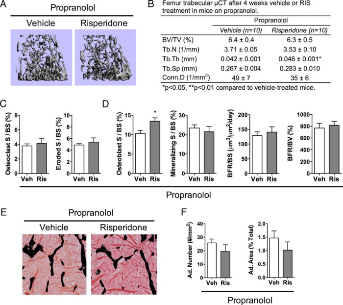
PRO prevented trabecular bone loss from RIS. RIS or vehicle (Veh) was administered to mice that were also receiving PRO treatment for 4 weeks, and analyses were performed at 12 weeks of age. A and B, Representative images (A) and quantitative parameters from μCT (B) of the trabecular bone of the distal femur. BV/TV, Tb.N, Tb.Th, Tb.Sp, and Conn.D were measured at 10-μm resolution. C and D, Quantitative osteoclast (C) and osteoblast (D) parameters from histomorphometric analyses of the trabecular bone of the proximal tibia (S, surface; BS, bone surface; BFR, bone formation rate; BV, bone volume). E, Representative images from histomorphometric analyses in the proximal tibia, where black stain is mineral (Von Kossa) and adipocytes ghosts are white. F, Quantitative adipocyte (Ad.) measurements from the proximal tibia. n = 5 to 9. *, P < .05.

