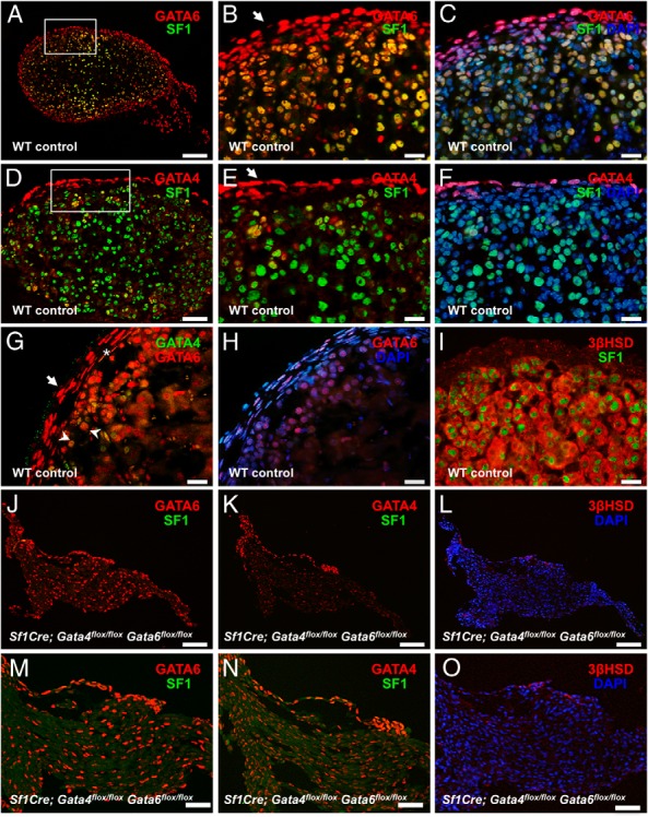
Figure 2.

Loss of steroidogenic cells in Sf1Cre;Gata4flox/floxGata6flox/flox adrenals. Representative sections from controls (A–I) and Sf1Cre;Gata4flox/floxGata6flox/flox (J–O) adrenals at E15.5 were stained for GATA6 (red) and SF1 (green) (A–C, J, and M); GATA4 (red) and SF1 (green) (D–F, K, and N); GATA6 (red) and GATA4 (green) (G); and 3βHSD (red) (I, L, and O) and SF1 (green) (I). DAPI (blue) was used as nuclear staining. In the control adrenals, most SF1-positive cells express GATA6 but not GATA4 (compare B and E). Only anterior capsular cells consistently express GATA4 (arrow in E), whereas most the capsular cells express GATA6 (arrows in B and G). G, A small subset of subcapsular cells (arrowheads) and rare capsular cells (asterisk) coexpresses both GATA factors. In J–O, note that steroidogenic (SF1- or 3βHSD-positive) cells are absent in the Sf1Cre;Gata4flox/floxGata6flox/flox adrenals. B and C, and E and F, are higher magnifications of A and D, respectively, and M–O are higher magnifications of J–L, respectively. Scale bars, 100 μm (A and J–L), 50 μm (D and M–O), and 20 μm (B, C, and E–I).
