Figure 4.
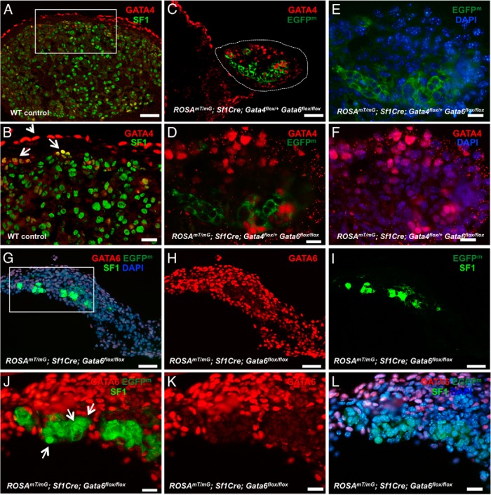
Adrenocortical development is severely impaired in the absence of GATA6. A–F, Representative sections from control (A and B) and ROSAmT/mG; Sf1Cre;Gata4flox/+Gata6flox/flox (C–F) adrenals at E15.5 were stained for SF1 (bright green) (A and B) and GATA4 (red) (A–D and F). Cells that underwent Sf1Cre-mediated recombination are traced by membrane EGFP (EGFPm) (dark green). In B note that GATA4 expression is mostly limited to capsular cells (arrowhead), with rare cortical cells expressing the protein (arrows). In contrast, numerous cortical cells are GATA4-positive in the ROSAmT/mG; Sf1Cre;Gata4flox/+Gata6flox/flox adrenal (C, D, and F; encircled in C). In C–E only some EGFP-positive cells express GATA4 (see also Supplemental Figure 4). G–L, A limited number of steroidogenic cells is present in the ROSAmT/mG; Sf1Cre;Gata6flox/flox adrenals. Adrenal sections of ROSAmT/mG; Sf1Cre;Gata6flox/flox at E15.5 were stained with antibodies against GATA6 (red; G, H, and J–L) and SF1 (bright green) (G, I, J, and L). EGFPm expression (dark green) traces SF1Cre-mediated excision (G, I, J, and L). In J, arrows point to SF1-positive nuclei that are strictly confined to the EGFPm-positive/GATA6-negative cells. J–L, Higher magnifications of a rectangular area in G. Scale bars, 50 μm (A, C, and G–I) and 20 μm (B, D–F, and J–L).

