
Figure 1.
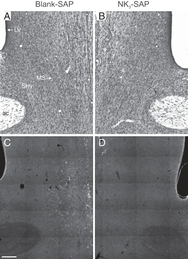
Representative coronal photomicrographs of the septum from rats injected with blank-SAP (left) or NK3-SAP (right). Cresyl violet–stained sections (A and B) show no qualitative change in the Nissl architecture between blank-SAP and NK3-SAP rats. In contrast, immunohistochemical studies (C and D) reveal a marked reduction in NK3R-ir cells in rats with NK3-SAP injections. Sections are shown as mirror images with landmarks labeled in A: ac, anterior commissure; LV, lateral ventricle; MS, medial septum, SHy, septohypothalamic nucleus. Scale bar corresponds to 250 μm.