
Fig. (4).
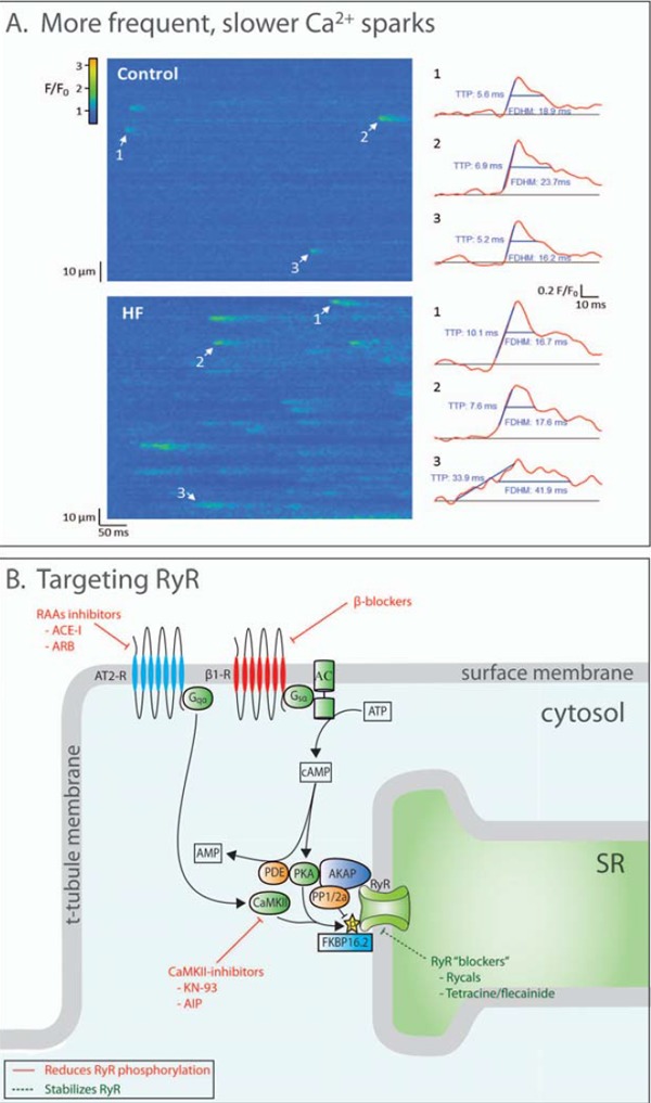
Altered Ca2+ sparks in failing cardiomyocytes and therapeutic targets of RyR activity. A: Line scan confocal imaging of failing cardiomyocytes (post-infarction mouse) reveals more frequent and slower Ca2+ sparks (temporal profiles of indicated sparks shown at right; reproduced from [6], with permission). Thus, disrupted RyR function in heart failure promotes SR Ca2+ leak and dyssynchronous Ca2+ release. B: RyR function is regulated by a large protein complex. Phosphorylation (by PKA and CaMKII) and dephosphorylation (by protein phosphatase 1 or 2a) are important regulatory pathways. Strategies to inhibit RyR phosphorylation, such as CaMKII inhibitors, are demonstrated to reduce SR Ca2+ leak. RyRs “blockers” such as rycals are an alternative approach to reducing leak.