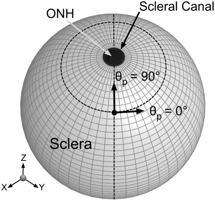
Figure 2.

An idealized FE model of the posterior hemisphere of an eye. The fiber orientation was defined such that θp = 0° represents the circumferential orientation (i.e. preferred fiber orientation tangent to the scleral canal boundary) and θp = 90° the meridional orientation (i.e. preferred fiber orientation perpendicular to the scleral canal boundary). The ONH is considered as the posterior hemisphere's pole, which includes the lamina cribrosa and retinal ganglion cell axons.
