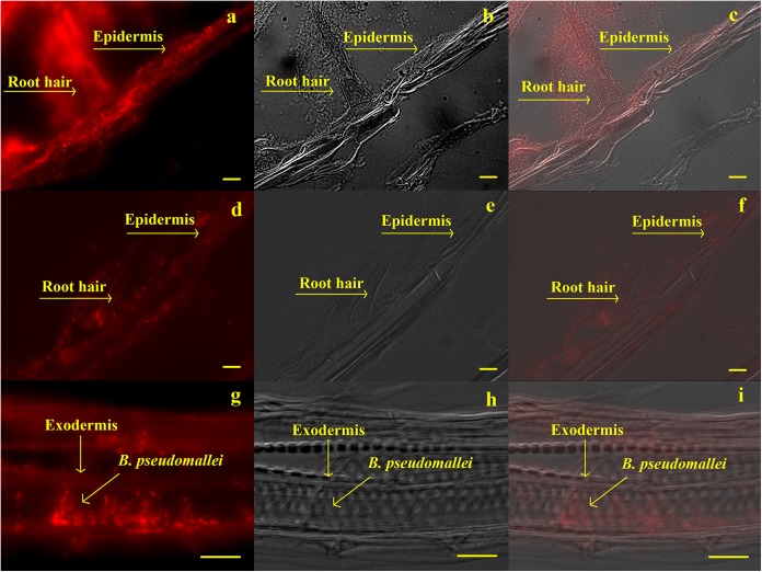
FIG 5.
MAb MCA2823 immunofluorescence results. Panels a, d, and g show B. pseudomallei labeled by IFA; panels b, e, and h are pictures of the same field taken by Nomarski interference contrast; and panels c, f, and i are combined images. A biofilm of B. pseudomallei can be seen on the epidermis and root hairs in panels a, b, and c. B. pseudomallei can also be seen inside the plant root hair in panels d, e, and f, and inside the exodermis in panels g, h, and i. Bars, 10 μm.

