
Fig. 3.
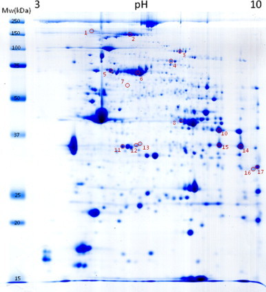
Coomassie blue staining of one representative 2D gel. The numbers and positions of the 17 selected spots identified by MS/MS correspond to those found as consistently increased on the old group (RMI>1.3).
Official websites use .gov
A
.gov website belongs to an official
government organization in the United States.
Secure .gov websites use HTTPS
A lock (
) or https:// means you've safely
connected to the .gov website. Share sensitive
information only on official, secure websites.
Coomassie blue staining of one representative 2D gel. The numbers and positions of the 17 selected spots identified by MS/MS correspond to those found as consistently increased on the old group (RMI>1.3).