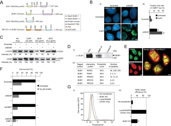
FIG 5.
Bub1 NHEJ is conserved in mammalian cells. (A) Schematic showing the conserved domain organization and length (amino acids) of Bub1 and Bub2 in budding yeast and humans. Colored boxes indicate the homology of the domain regions. (B) Accumulation (I) and quantitative (II) analysis of 53BP1 foci (green) formation in U2OS cells transfected with BUB1 shRNA in the presence and absence of cisplatin (CisPt). DAPI (blue) was used for nuclear staining and visualization of 53BP1 foci. The scale bar equals 5 μm. (C) Immunoblot analysis of H2AX phosphorylation levels in shBUB1 and control-depleted cells in the presence and absence of the indicated concentration of cisplatin (CisPt), bleomycin (BLM), or neocarzostatin (NCS) using an anti-γ-H2AX antibody. Epidermal growth factor receptor-binding protein (GRB2) probed with an anti-GRB2 antibody was used as the loading control (LC). (D) Immunoblot analyses (I) of 53BP1 in the input whole-cell lysate (WCL) and anti-53BP1 immunoprecipitates (IP) using anti-BUB1 antibody in human U2OS cells. The molecular mass of the marker protein by SDS-PAGE is indicated. NHEJ proteins copurified from the endogenous BUB1 (II) using anti-BUB1 antibody (peptides from three independent replicates shown) in human U2OS cells were identified at high confidence (99%) by MS/MS. (E) Immunofluorescent images showing the 53BP1 (green) colocalization with BUB1 (red) in the human U2OS cells. Cells were immunostained with anti-53BP1 and anti-BUB1 antibodies. The scale bar equals 8 μm. (F) Clonogenic survival of control shRNA and BUB1- and 53BP1-depleted U2OS cells treated with cisplatin (CisPt; 32 μM). Asterisks indicate significantly reduced (P ≤ 0.05, Student's t test) cell survival of BUB1 53BP1 double knockdowns compared to their corresponding single knockdowns. (G) FACS-sorted positive GFP cells (I), indicating NHEJ repair efficiency (II) in U2OS cells transfected with the empty LentiCRISPR vector (blue) or the BUB1 CRISPR knockout (KO [green]). U2OS cells not transfected with the indicated knockout or vector (red) served as a control. Fluorescence values are in arbitrary units. Data (B and G) are represented as the mean ± SEM (n ≥ 3); asterisks indicate significant (P ≤ 0.05, Student's t test) difference between the indicated knockdowns or knockouts versus scrambled shRNA or LentiCRISPR control.

