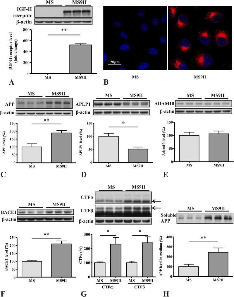
FIG 1.
(A and B) Western blotting (A) and immunofluorescence staining (B) validating increased levels and expression of IGF-II receptor in MS9II versus MS cells. (C to E) Immunoblots and respective histograms showing increased levels of APP holoprotein (C), decreased levels of homologous APLP1 protein (D), and unaltered levels of ADAM10 (E) in IGF-II receptor-overexpressing MS9II cells. (F to H) Immunoblots and respective histograms showing increased levels of BACE1 (F), APP-CTFs (CTF-α and CTF-β) (G), and soluble APP fragments (sAPPα and sAPPβ) (H) in MS9II cells versus MS cells. All Western blots were reprobed with β-actin antibody to monitor protein loading, and the values, expressed as means ± SEM, were from 3 or 4 independent experiments. Data were analyzed using Student's t test. *, P < 0.05; **, P < 0.01. Scale bar, 20 μm.

