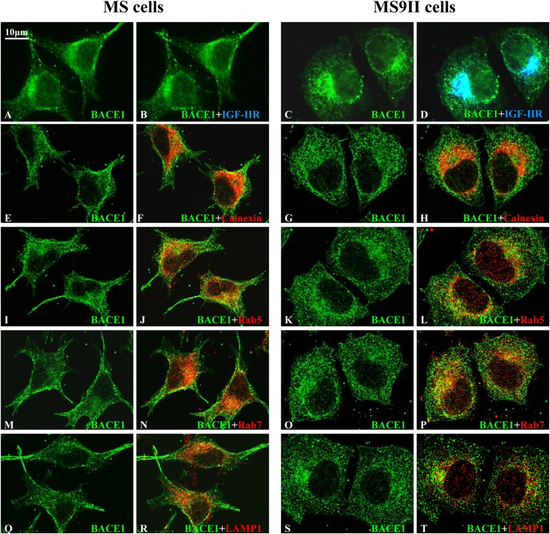
FIG 5.
(A to D) Representative immunofluorescence images of MS and MS9II cells showing localization of BACE1 with IGF-II receptor. Note the localization of a subset of BACE1 with IGF-II receptor in MS9II cells. (E to T) Confocal images of MS and MS9II cells depicting localization of BACE1 (E, G, I, K, M, O, Q, and S) in calnexin-labeled ER (F and H), Rab5-labeled early endosomes (J and L), Rab7-labeled late endosomes (N and P), and LAMP1-labeled lysosomes (R and T). IGF-II receptor, as expected, was not detected in MS cells. BACE1 immunoreactivity was more evenly distributed in the ER, endosomes, and lysosomes in MS cells than in MS9II cells.

