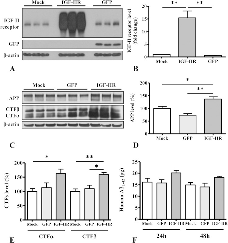
FIG 9.
(A and B) Immunoblots (A) and respective histogram (B) showing levels of IGF-II receptor in human SK-N-AS neuroblastoma cells following infection with adenoviral constructs for the IGF-II receptor and green fluorescent protein (GFP) versus mock-infected cells. (C to E) Immunoblots (C) and respective histograms showing increased levels of APP (D) and APP-CTFs (E) in IGF-II receptor overexpressing SK-N-AS neuroblastoma cells compared to levels in mock-infected and uninfected control cells. (F) Histogram showing levels of secretory Aβ1–42 in the conditioned media of human SK-N-AS neuroblastoma cells transduced with the IGF-II receptor or GFP compared to that of mock-infected cells at 24 and 48 h after infection. All Western blots were reprobed with β-actin antibody to monitor protein loading, and the values, expressed as means ± SEM, were from 2 or 3 independent experiments. All data were analyzed using Student's t test. *, P < 0.05; **, P < 0.01.

