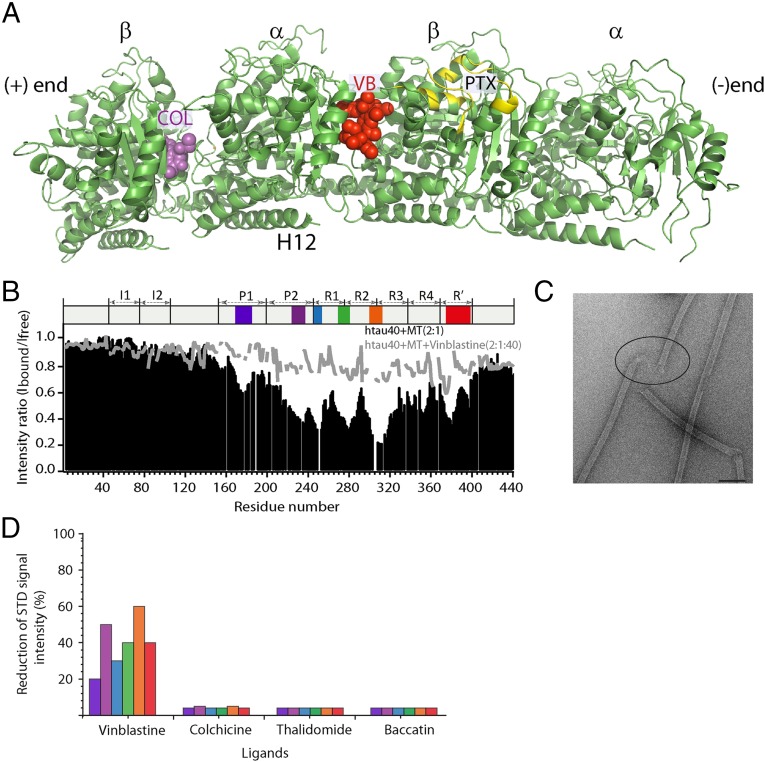
Fig. 2.
Tau competes with vinblastine for binding to tubulin. (A) Location of vinblastine (VB), colchicine (COL), and paclitaxel (PTX) on the 3D structure of the (Tc)2R complex (PDB ID code 1Z2B; ref. 31). Bound colchicine and vinblastine are shown by using the sphere model. Residues involved in paclitaxel binding (54) are highlighted in yellow. (B) Vinblastine competes with four-repeat Tau for binding to microtubules. Black bars represent the NMR line broadening observed in four-repeat Tau upon addition of microtubules (molar ratio of 2:1), whereas the gray line shows the corresponding values after addition of a 20-fold excess (with respect to four-repeat Tau) of vinblastine. (C) Electron micrograph of paclitaxel-stabilized microtubules after addition of vinblastine. The concentrations of Tau, microtubules and vinblastine were 10, 20, and 400 μM, respectively, in 50 mM sodium phosphate buffer, pH 6.8. (D) Competition between Tau fragments and vinblastine for binding to unpolymerized tubulin as probed by saturation-transfer difference NMR (SI Appendix, Figs. S4 and S5). Each of the six Tau fragments Tau(162-188) (blue), Tau(211-242) (purple), Tau(239-267) (light blue), Tau(265-290) (green), Tau(296-321) (orange), and Tau(368-402) (red) overlaps with one of the microtubule-binding motifs (Fig. 1 and SI Appendix, Fig. S1).

