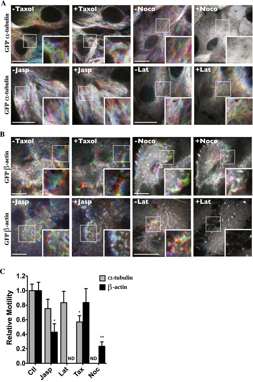
Fig. S6.
Microfilament motility is differentially affected by chemicals that interfere with microfilament stability. (A and B) Live-cell spinning-disk images of GFP-tagged α-tubulin (A) and GFP-tagged β-actin (B) were collected before and after the addition of taxol (10 µM), nocodazole (10 µM) (Noco), jasplakinolide (0.4 µM) (Jasp), or latrunculin B (2.5 µM) (Lat) to LLC-PK1 cells and are color-coded as described in Materials and Methods. Images depicting motility during a 6-min timeframe prechallenge and 14 min postchallenge indicate that taxol interferes moderately with MT motility, and nocodazole and jasplakinolide appear to reduce actin motility. (Scale bars: 10 µm.) Representative images from three independent experiments are shown. (C) Quantification of microtubule and actin motility. Data were gathered from three independent experiments. *P < 0.05, **P < 0.01 vs. control. ND, not determined.

