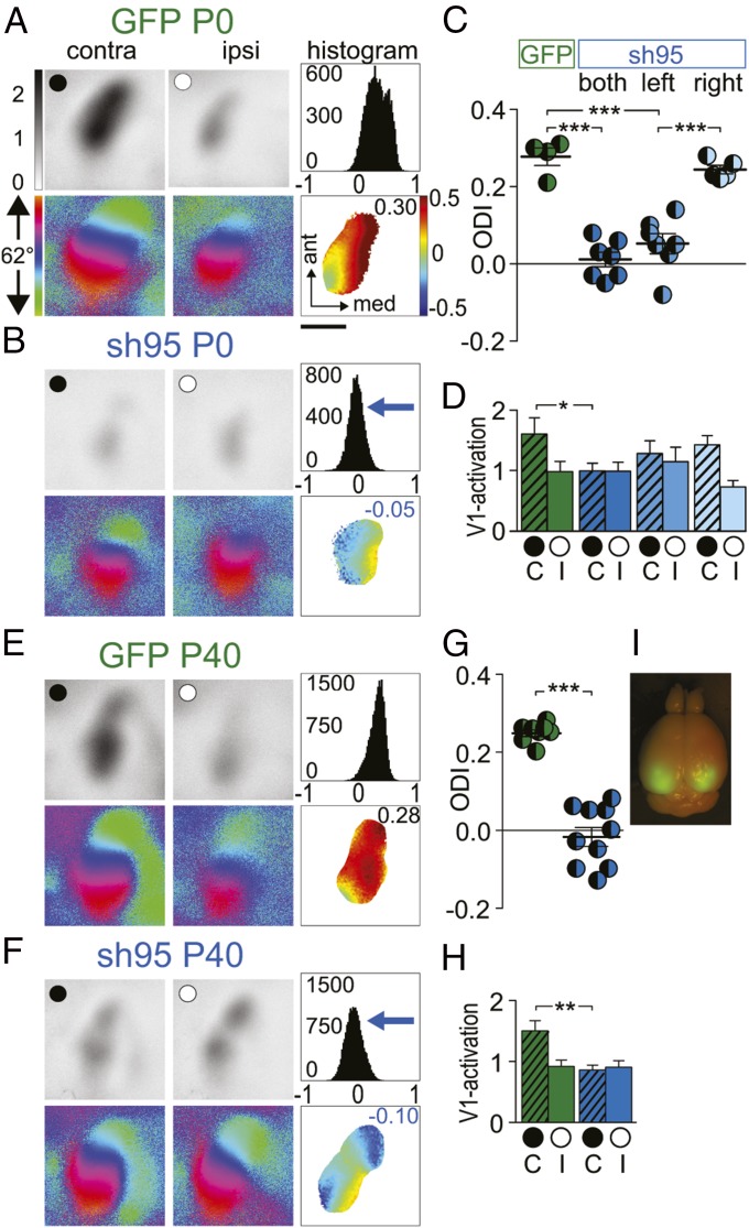
Fig. 6.
PSD-95 controls the closure of the critical period for juvenile ocular dominance plasticity. Optically imaged activity maps (A, B, E, and F) and their quantification (C, D, G, and H) in V1 of AAV-GFP and AAV-sh95 mice transduced at P0 (A–D) and P40 (E–H). V1 recordings after a 4-d MD at >P80. Data displayed as in Fig. 1. (Scale bar, 1 mm.) ODIs (C and G) and V1 activation (D and H) after a 4-d MD in AAV-GFP–transduced mice at P0 (C and D) or P40 (G and H); AAV-sh95 transduced bilaterally at P0 (both; A, C, and D) or P40 (E, G, and H) or at P0 only contra- (left; C and D) or ipsilateral to the deprived eye (right; C and D) (GFP P0 vs. sh95 P0 right hemisphere, P = 0.20). (D and H) V1 activation elicited by stimulation of the contralateral (C) or ipsilateral (I) eye. (I) GFP fluorescence was illuminated in a perfusion fixed brain, which was transduced on P40 with AAV-sh95 and analyzed on P80 (see also confocal microscopy of slices in Fig. S6). *P < 0.05; **P < 0.01; ***P < 0.001. Values in Table S1.

