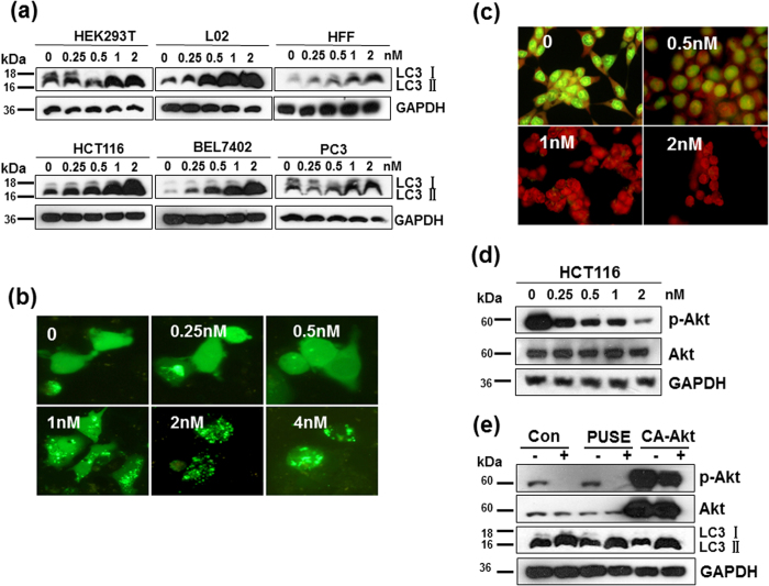
Figure 4. CTAB-coated GNRs induced autophagy in cells.
Cells were treated with 0.25–2 nM CTAB-coated GNRs for 24 h. The autophagy of HCT116 cells induced by CTAB-coated GNRs was Akt independent. (a) Western blot analysis of LC3 protein levels in cancer cell lines as well as in immortalized nonmalignant cell lines. Cropping lines are used in the figure. Full-length blots are presented in Supplementary Figure 2. The gels have been run under the same experimental conditions. (b) Representative GFP-LC3 fluorescent punctate dot images in HCT116. (c) Detection of acidic vesicular organelles with acridine orange staining. (d) Western blot analysis of Akt in cells treated with 0.25–2 nM CTAB-coated GNRs for 24 h. Cropping lines are used in the figure. Full-length blots are presented in Supplementary Figure 2. The gels have been run under the same experimental conditions. (e) Cells transfected with vehicle plasmid (pUSE) or constitutively active Akt (CA-Akt) were incubated with or without 2 nM CTAB-coated GNRs for 24 h and then analyzed for LC3 and Akt by Western blot. Cropping lines are used in the figure. Full-length blots are presented in Supplementary Figure 2. The gels have been run under the same experimental conditions.

