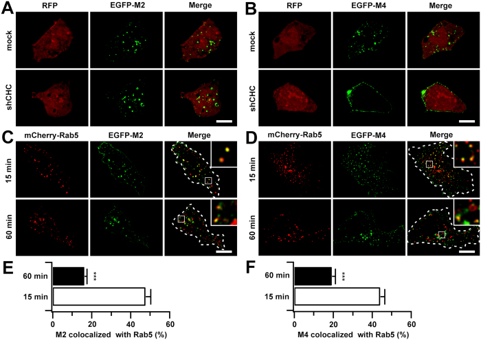
Figure 2. M2 and M4 mAChRs were transported to Rab5-positive early endosomes via different endocytic pathways.
(A,B) HEK293 cells expressing EGFP-M2 (A) or EGFP-M4 (B) receptors were transfected with mock or CHC shRNA and were assessed for CCh-stimulated internalization by confocal microscopy. Shown only are cells treated for 60 min of CCh (100 μM). (C,D) HEK293 cells were co-transfected with EGFP-M2 (C) or EGFP-M4 (D) and mCherry-Rab5. Representative confocal images show the localization of internalized M2 or M4 receptors with Rab5 following 15 or 60 min stimulation of CCh (100 μM). Shown only are cells treated for various time of CCh. Cell contours were outlined and insets showed the magnified boxed regions. Scale bars, 10 μm. (E,F) The levels of colocalization of EGFP-M2 (E) and EGFP-M4 (F) with Rab5 depicted in C and D were determined by Manders coefficient as described in ′′Methods′′. ***p < 0.001.

