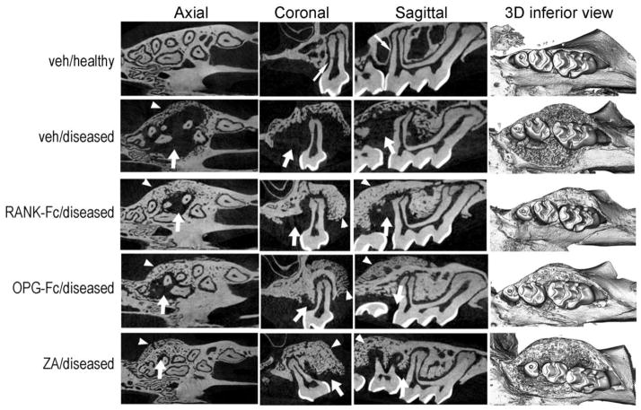
Figure 1.
Radiographic changes of the maxillary alveolar ridge. Representative axial, coronal and sagittal μCT slices and three-dimensional views of the maxillary molars of healthy (veh) or diseased site from veh, RANK-Fc, OPG-Fc and ZA treated animals are shown. Thin arrows point to the lamina dura around the roots of the veh/healthy animals. Thick arrows point to areas of osteolysis and arrowheads to areas or bone expansion in the diseased site of veh, RANK-Fc, OPG-Fc and ZA treated mice.

