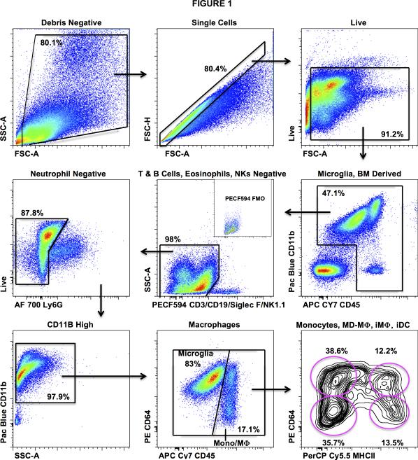
Figure 1. Gating strategy for the differentiation of microglia from monocytic cells.
Cells were isolated from enzymatically digested, myelin depleted, mouse brains. After the exclusions of doublets and debris, immune cells were identified by CD45 staining. Live cells were separated from dead cells using an amine reactive dye. Lymphocytes, eosinophils, and NK cells were excluded using a common channel for CD3/CD19, Siglec F, and NK1.1 respectively. Neutrophils were identified and gated out using surface staining for Ly6G. Microglia (CD3-CD19−NK1.1−SiglecF−Ly6G−CD11b+CD45Lo) were then differentiated from infiltrating monocytes and macrophages (CD3−CD19−NK1.1−SiglecF−Ly6G−CD11b+CD45Hi) by differential expression of CD64 and CD45. Backgating based on the differential forward scatter characteristics and CD11b expression aided identification of these populations. Monocytes (CD64−MHCII−), early monocyte-derived (CD64+MHCII−) macrophages (MD-Mϕ), inflammatory (CD64+MHCII+) macrophages (iMϕ), and inflammatory (CD64−MHCII+) dendritic cells (iDC) were then identified via overlapping expression patterns of CD64 and MHCII.

