FIGURE 2.

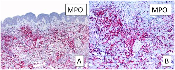
Myeloperoxidase stain for myeloid cells. A, Strong myeloperoxidase positivity reveals the presence of cells from a myeloid origin (original magnification, 10X). B, Higher magnification of A (40X). MPO: myeloperoxidase.

Myeloperoxidase stain for myeloid cells. A, Strong myeloperoxidase positivity reveals the presence of cells from a myeloid origin (original magnification, 10X). B, Higher magnification of A (40X). MPO: myeloperoxidase.