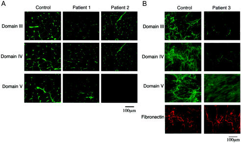
Figure 2.
Immunostaining of perlecan in muscle tissues and cultured fibroblasts. A, Muscle tissue from patients 1 and 2 and from an unaffected control subject, stained with domain-specific anti-perlecan antibodies as described elsewhere (Arikawa-Hirasawa et al. 2001, 2002). In patient 1, antibodies to domains III–V stained the basal lamina of the muscle, whereas, in patient 2, domain V staining was absent. The staining in muscle tissue of patient 1 is significantly reduced. B, Cultured fibroblasts from patient 3 stained with domain-specific anti-perlecan antibodies and with anti-fibronectin polyclonal antibodies. Domains III–V stained the extracellular matrix at significantly reduced levels compared to control fibroblasts, whereas fibronectin stained strongly in both.

