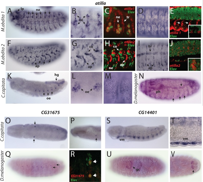
Fig. 5.
Embryonic tissue-specificities of atilla, CG14401, and CG31675 genes. (A–N) atilla genes. (A–E) Megaselia abdita atilla1. (A, B) Lateral views showing expression in the lateral sensory neurons (ne) and the larval photoreceptors (lp). (C) Fluorescent double staining showing atilla1 (red) and Elav protein (green) distribution on the lateral sensory neurons (ne). atilla1 expression is detected in both neurons and trachea (tr). (D) Expression in the dorsal epidermis. (E) Ventral view of atilla1—Elav double staining showing atilla1 expression in the Elav-negative glial cells associated with the exiting nerves (arrows) and within the VNC (inset). (F–J) Megaselia abdita atilla2. (F, G) Lateral views displaying expression in the muscle apodemes (ap) and the chordotonal organs (co). (H) Fluorescent staining of atilla2 (red) and Elav protein (green) on the lateral sensory organs. atilla2 transcripts are found both in nonneuronal components of the chordotonal organs (co) and in few Elav positive sensory neurons on the ventral side (ne). (I) Expression in the dorsal heart (hr) and apodemes (ap). (J) atilla2 is expressed in Elav positive cells in the VNC (magnified in inset). (K–M) Ceratitis capitata atilla. (K, L) Lateral views showing expression in the hindgut (hg) and the oenocytes (oe). (M) Late embryos show expression in the epidermis. (N) Drosophila melanogaster lateral view. atilla is expressed in the epidermis, trachea (arrows), and pharynx (ph). (O–R) CG31675 orthologs. (O, P) Ventral (O) and lateral (P) views of C. capitata embryo at the extended germband stage showing labeling of unidentified groups of cells posterior to the head (arrow). (Q, R) Drosophila melanogaster CG31675. (Q) Lateral view showing expression in the dorsal sensory organs. (R) CG31675 (red) is expressed in Elav-positive (green) lateral neurons (arrows). (S–V) CG14401 orthologs. (S, T) Ceratitis capitata. (S) Lateral view showing CG14401 expression in the ventral longitudinal muscles (vm). (T) Magnified view of the ventral muscle, seen from a ventrolateral perspective. (U, V). Drosophila melanogaster CG14401. (U) Ventral view showing expression in the garland cells (gc). (V) Dorsal view showing expression in a subset of cells associated with the posterior spiracles (arrow).

