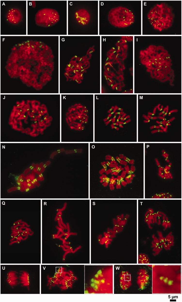
Fig. 2.
Organization of CenH3-containing domains on mitotic chromosomes at different levels of chromatin compaction. (A and B) Immunodetection of CenH3 in the nuclei of Pisum sativum (2n = 14) (A) and Vicia faba (2n = 12) (B). Note that the number of CenH3 signals corresponds to the number of chromosomes. (C–W) Immunodetection of CenH3 at different levels of chromatin compaction during mitosis. (C–F) Chromosomes at the early stages of chromatin condensation in P. sativum (C), V. pannonica (D), V. sativa (E), V. faba (F), P. sativum (G), P. fulvum (H), V. pannonica (I), V. faba (J), and V. sativa (K). Note that the low-condensed chromosomes in Pisum display the beads on a string pattern while those in Vicia possess a single uninterrupted CenH3 signal. (L and M) Metaphase chromosomes in P. sativum (L) and P. fulvum (M) displaying mostly ribbon-like patterns of CenH3 distribution. (N) Metaphase chromosomes in La. sylvestris showing either beads on a string or ribbon-like patterns of CenH3 distribution. (O) Metaphase chromosomes in La. sativus displaying the beads on a string pattern. (P and S) Metaphase chromosomes in V. sativa (P), V. pannonica (Q), V. faba (R), and Lens culinaris (S) showing single dot-like CenH3 signals. (T) Anaphase of chromosomes in P. fulvum, some of which show a ribbon-like pattern of CenH3 signal. (U) Anaphase chromosomes in Le. culinaris with single dot-like signals of CenH3 at each centromere. (V and W) anaphase (V) and telophase (W) chromosomes in La. sativus showing the beads on a string pattern. Insets show 5 × magnifications of the boxed regions. The immunodetection experiments were performed using squash preparations of synchronized root tip meristems fixed in 4% formaldehyde. CenH3 (green) was immunodetected with antibodies primarily raised to CenH3-1_PSat (P. sativum and P. fulvum), CenH3-2_PSat (La. sativus and La. sylvestris), CenH3-2_VF (V. faba, V. sativa and V. pannonica), and CenH3-2_LCul (Le. culinaris). Chromosomes were counterstained with DAPI (red). Bar = 5 µm.

