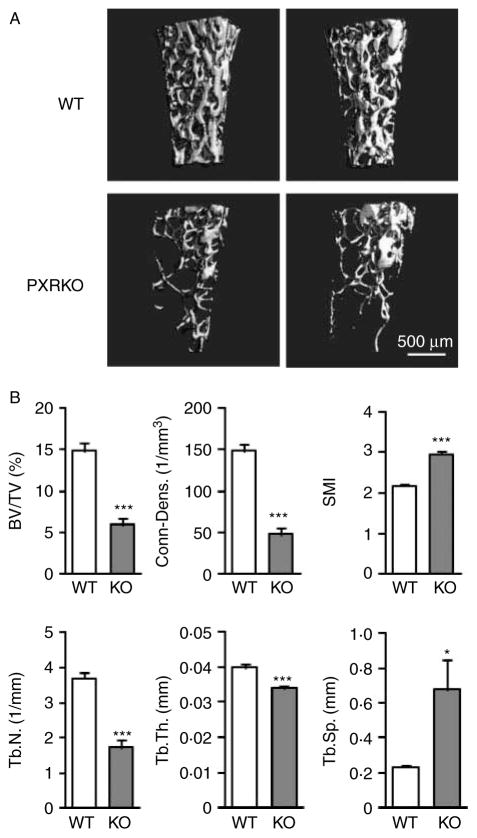
Figure 2.
Three-dimensional change in the trabecular bones of PXR knockout mice. (A) Representative three-dimensional micro-CT images of the femoral trabecular bones are shown. Upper panels: wild-type (WT) mice. Lower panels: PXR knockout mice (PXRKO). (B) Microstructural parameters (BV/TV, three-dimensional bone volume fraction per tissue volume; Conn-Dens., connectivity density; SMI, structure model index; Tb.N., trabecular number; Tb.Th., trabecular thickness; Tb.Sp., trabecular separation) of femoral trabecular bones of wild-type (WT; n=8) and PXR knockout (KO; n=8) mice derived from micro-CT analysis are shown. ***P<0·001; *P<0·05.

