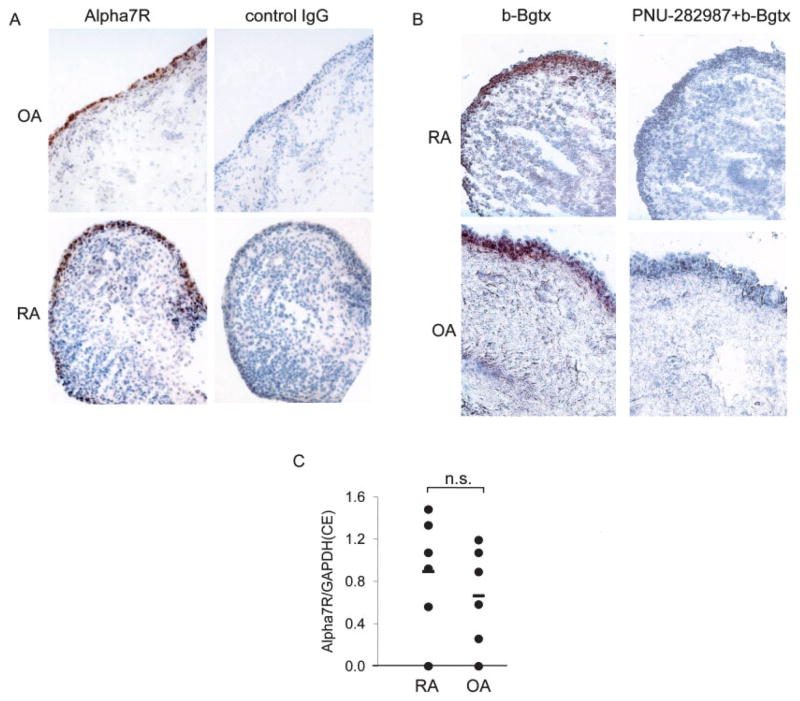
Figure 1.
Expression of the α7 receptor subunit (α7R) in synovial tissue from rheumatoid arthritis (RA) and osteoarthritis (OA) patients. A and B, Immunohistochemistry of α7R in RA and OA synovial tissue was performed with an anti-α7R monoclonal antibody or control IgG (A) or with biotin-labeled bungarotoxin (b-Bgtx), with or without PNU-282,987, a selective agonist of α7R (B), as described in Materials and Methods. Staining for the receptor was highest in the intimal lining. Representative serial sections from an RA and an OA patient (of 3 RA and 3 OA samples examined) are shown. C, Expression of α7R mRNA in 6 RA and 6 OA synovial tissue samples was not significantly different (NS). Values are the ratio of α7R expression to GAPDH expression. Horizontal bars show the mean. CE = relative cell equivalents (see Materials and Methods for details).

