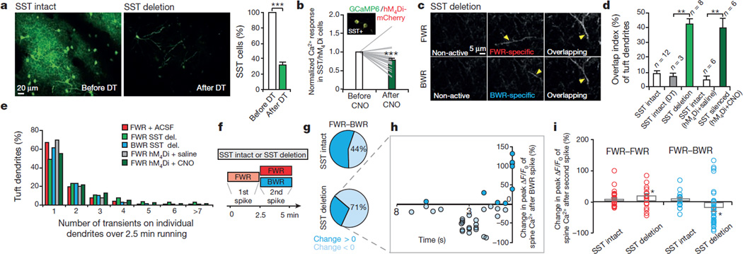
Figure 4. Disrupting cortical inhibition alters branch-specific Ca2+ spikes and synaptic potentiation.
a, Two-photon images of SST-interneurons labelled with green fluorescent protein (GFP) in motor cortex before and 2 days after diphtheria toxin (DT) treatment. Loss of SST interneurons occurred within 2 days after diphtheria toxin treatment. b, Somatic Ca2+ activity was reduced after CNO in SST interneurons expressing hM4Di-mCherry/GCaMP6s (inset). c, Two-photon images of tuft dendrites exhibiting spikes during FWR and BWR after SST deletion. d, SST deletion or silencing significantly increased the percentage of tuft dendrites exhibiting Ca2+ spikes during both FWR and BWR (SST/DTR: P = 0.0007; SST/hM4Di: P = 0.002). e, Inactivating SST interneurons did not affect the number of Ca2+ spikes generated during FWR as compared to control mice (P> 0.05). More than 200 Ca2+ spikes were measured per condition. del., deletion. f, Training protocol to test the effect of disrupting branch-specific Ca2+ spikes on potentiated spine Ca2+ transients in control and SST-deleted mice. g, Pie charts showing 44% and 71% of FWR-potentiated spines were depotentiated during BWR in control and SST-deleted mice, respectively. h, Spines potentiated during FWR were depotentiated when active <5 s before BWR-induced Ca2+ spikes in SST-deleted mice. i, Summary of the effect of FWR or BWR on previously potentiated spines in control and SST-deleted mice. Data are mean ± s.e.m. *P < 0.05, **P < 0.01, ***P < 0.001, paired t-test (a, d, i) and Wilcoxon matched-pairs signed rank test (b, i). Statistical details in Methods.

