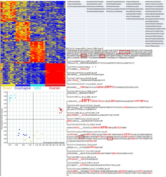
Figure 5.
Specificity of immunosignatures (left) and mapping of Glioblastoma multiformae-specific peptide motifs to translations of RNA libraries from tumor libraries: Upper left: heatmap demonstrates the differences in immunosignatures among four different cancers, stage III breast cancer, esophageal adenocarcinoma, Glioblastoma multiformae (GBM, a stage IV astrocytoma), and stage IV (a and B) ovarian cancer. Peptide intensities are shown on the Y-axis, and patients on the X-axis. Peptides and patients are grouped by hierarchical clustering based on the data, indicating which patient samples show reactivity with which peptides. the principal components analysis map (PCA) immediately below the heatmap shows further grouping by inter- and intragroup variances. Top right: panel lists peptides reactive in GBM patients. Below right: panel shows the alignment of sequences from a translated RNA tumor library obtained from GBM patients, with the alignment of GBM-specific immunosignature peptides and the particular gene or gene constituents (if a translocation) preceding each line. The red letters indicate the motifs found in the GBM-specific peptides found using the 330,000-peptide (high-density) library, with the height indicating the amount of conservation in that position. The larger the letter, the more often that amino acid was found at that specific position in the translated library. Squares indicate that a motif was also found using a 10,000-peptide (low-density) printed peptide microarray.

