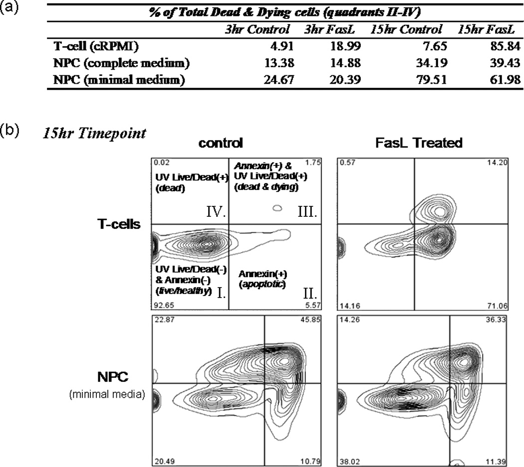
Fig. 2.

FasL does not induce cell death in NPCs. T cells and NPCs were treated with FasL (200 ng/ml) plus an enhancer (4 g/ml) for 3 and 15 hr at 37°C before UV blue Live/Dead dye and annexin Alexafluor 647 staining. Cells were subsequently analyzed by flow cytometry. a: Summary of results, where T cells or NPCs were placed in either plain media or media plus FasL for 3 or 15 hr. b: Readout from the 15-hr treatment as gated using Flow Jo software. As illustrated in b, quadrant I represents cells living healthy cells, which stain negative for either dye; quadrant II represents early apoptotic cells, which are positive for annexin; quadrant III represents dead and dying cells, which stain positively for both annexin and UV Live/Dead dye; and quadrant IV includes only necrotic cells (i.e., positive for only UV Live/Dead dye). A dramatic increase in the amount of apoptotic and/or necrotic cells (i.e., UV and/or annexin+ cells) was observed in T cells treated with FasL for 3 or 15 hr (top row of a and top plates in b). Compared with controls, no effect on NPCs was observed after 3 or 15 hr of FasL treatment in complete neurobasal medium supplemented with EGF and FGF (middle row of a). No induction of death was observed in NPCs treated for 3 hr with FasL in minimal medium compared with control cells (last row of a). A slight decrease (approximately 18%) in the total dead and dying cells was observed in NPCs treated with FasL for 15 hr compared with controls (bottom plates in b).
