
Fig. 5.
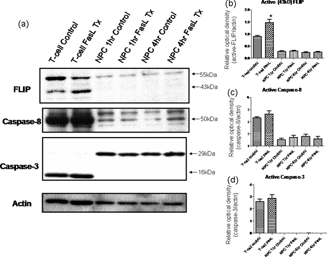
a–d: FasL does not influence FLIP or caspase levels in NPCs. Western blot results for probing the signaling cascades involved in Fas activation. Cell lysates obtained using NP-40 after various treatment periods of 200 ng/ml FasL + 4 µg/ml ą-flag (enhancer). The blot was first probed using an anti-FLIP antibody; then, the blot was consecutively stripped and reprobed for caspase-8, caspase-3, and finally β-actin. Secondary antibodies were HRP conjugated and visualized using ECL development reagents. FLIP levels remain unchanged in NPCs after FasL treatment; NPCs express low levels of caspase-8, and expression is not changed with FasL treatment at various time points; the levels of procaspase-3 (~29 kDa) remain unchanged. In contrast to the casde for T cells, the activated (cleaved) form of caspase-3 (~16 kDa) is not produced in NPCs after FasL treatment at various time points. *P < 0.05.