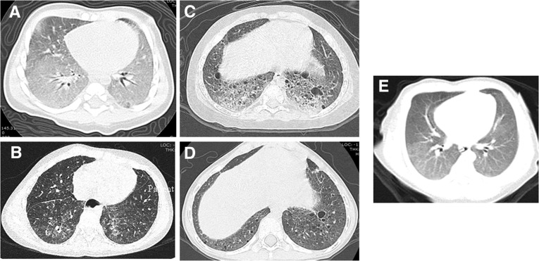
Figure 1.
Initial and most recent computed tomography (CT) scan for each patient. (A) Patient 1 initial CT scan with diffuse ground-glass opacities and septal thickening. (B) Patient 1 recent CT scan with nonspecific interstitial pneumonia findings, diffuse ground-glass opacities, and bronchiectasis. (C) Patient 2 initial CT scan with marked interstitial prominence, cystic change, and interstitial/alveolar infiltrate. (D) Patient 2 recent CT scan with interstitial prominence and increased number of parenchymal and subpleural cysts. (E) Patient 3 only CT scan with diffuse ground-glass opacities.

