Fig. 2.
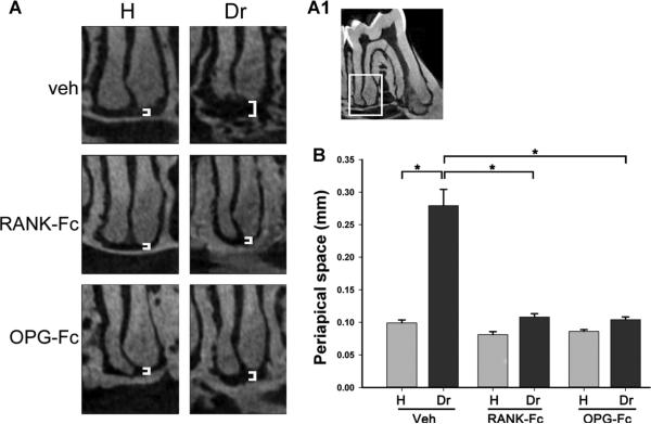
Changes in the periapical area of molars from animals treated with Veh, RANK-Fc, or OPG-Fc. (A) μCT images of the periapical area of the first molar distal root of healthy and drilled sites in animals treated with Veh, RANK-Fc, or OPG-Fc. (A1) The periapical area of the first molar distal root, as depicted in A. (B) Quantification of periapical space at the distal root of the first molar. *Statistically significantly different, p < 0.001. Veh = vehicle; RANK = receptor activator of NF-kB; Fc = fragment crystallizable; IgG = immunoglobulin G; RANK-Fc = extracellular domain of RANK fused to the Fc portion of IgG; OPG = osteoprotegerin; RANKL = RANK ligand; OPG-Fc = RANKL-binding domains of OPG linked to the Fc portion of IgG; μCT = micro–computed tomography.

