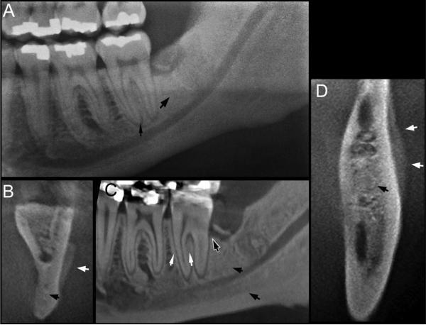
Fig. 5.
Panoramic (A), coronal (B), sagittal (C), and axial (D) CBCT sections through the left mandibular alveolar ridge of a patient with ONJ treated with denosumab. Thin white arrows point to the thickened lamina dura, thick white arrows to periosteal bone deposition, thin black arrow to widened periapical PDL space, thick black arrows to increased bone density, and black and white arrow to loss of periodontal bone at the distal surface of the second left mandibular molar. CBCT = cone beam computed tomography; ONJ = osteonecrosis of the jaws; PDL = periodontal ligament.

