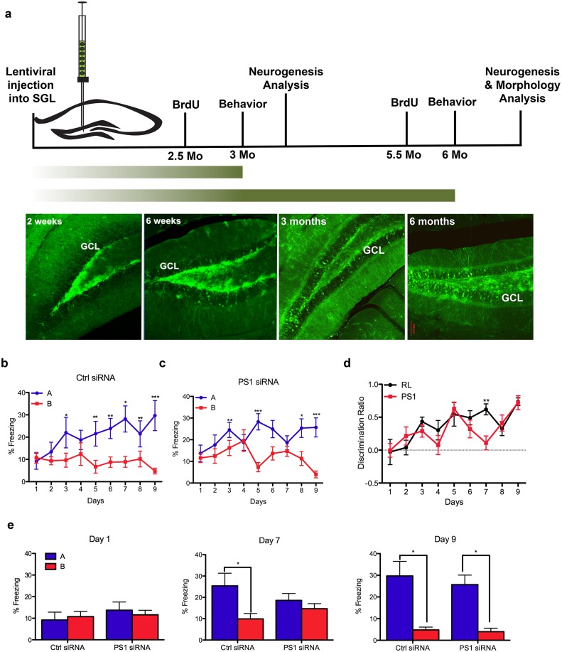
Fig 1. Impairments in pattern separation 3 months after PS1 knockdown in neural progenitor cells.
A. Experimental timeline. GFP expression in the dentate gyrus of the hippocampus at 2 weeks, 6 weeks, 3 and 6 months after lentivirus injection. Two weeks following injection, GFP-infected cells are located in the subgranular layer of the dentate gyrus. Migration of GFP+ cells into the granular cell layer is apparent at 6 weeks post injection, while at 3 and 6 months post-injection many GFP+ cells are incorporated in the granular cell layer. B-C Percentage of freezing in context A (shock) vs. context B (similar context) of mice 3 months following injection with either control (15) shRNA (B) or with PS1 shRNA (C) (two-way repeated measures (context and days) ANOVA, F(context)1,16 = 5.02, P<0.05; and F(context)1,18 = 6.18, P<0.05 respectively). Paired t-test, *p<0.05, **p<0.005. Error bars indicate ± SEM. D. Discrimination ratio [(A-B)/max(A,B)] between the control and the PS1 injected animals (two-way ANOVA [treatment and days] with repeated measures, F(treatment)1,153 = 1.38, P>0.05) Error bars indicate ±SEM. E. Post-hoc comparisons between contexts and treatments on single days during the recognition memory paradigm (paired t-test, *p<0.05).

