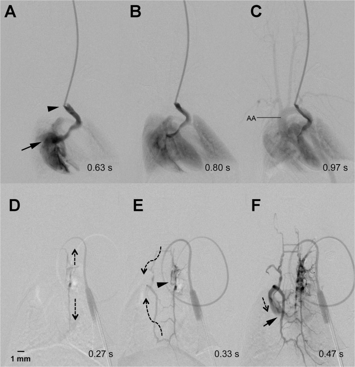
Fig 2. Collateral antegrade flow pattern.
The catheter tip (arrow head) is located within the jugular vein of the mouse. Contrast agent flows from the patent jugular vein into the right atrium (arrow) (A). From there it fills the lungs (B) and the aortic arch (AA) including the supraaortic arterial vessels (C). Contrast injection via the implanted mini-port one week later shows occlusion of the jugular vein (D). Instead, the contrast agent flowed (indicated by dotted arrows) via a “ladder-like” venous collateral network (E) with some delay and reduced contrast intensity into the right atrium (arrow) (F).

