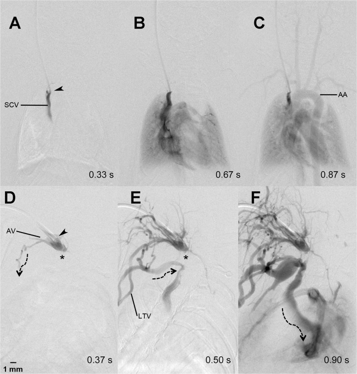
Fig 3. Collateral retrograde flow pattern.
Regular contrast agent flow through the heart, lung and aortic arch (AA) after contrast injection into a port-catheter implanted into the superior caval vein (SCV) (A-C). One week later, the catheter tip (arrow head) was dislocated slightly backwards into the confluens of the jugular and axillary vein (AV) and the SCV was thrombosed (*) (D). The contrast agent flowed (dotted arrows) into the proximal SCV via thoracic collaterals including the lateral thoracic vein (LTV) (E and F).

