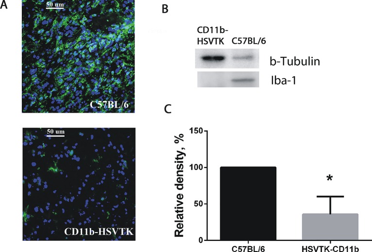
Fig 4. Microglia ablation in brain tumors using the CD11b-HSVTK/GCV system.
(A) Immunocytochemical detection of microglia in tumors developed in C57BL/6 and CD11b-HSVTK mice brains after local GCV administration. The tumors were generated by intracranial implantation of GL261 glioma cells. GCV was delivered to the tumor area through mini-osmotic pumps. Image shows the significant reduction of microglia in tumors developed in CD11b-HSVTK compared to C57BL/6 mice. Anti-Iba 1 antibody was used to detect microglial cells (green) and DAPI was used to detect all cell nuclei (blue). (B) Western blot detection of Iba-1 in tumors extracted from C57BL/6 and CD11b-HSVTK mice brains after the treatment with GCV. (C) The graph shows corresponding levels of Iba-1 protein expression in C57BL/6 and CD11b-HSVTK mice brain tumors after GCV administration determined by western blot. Mean ± S.E and significant difference from control (*) are shown (p < 0.05).

