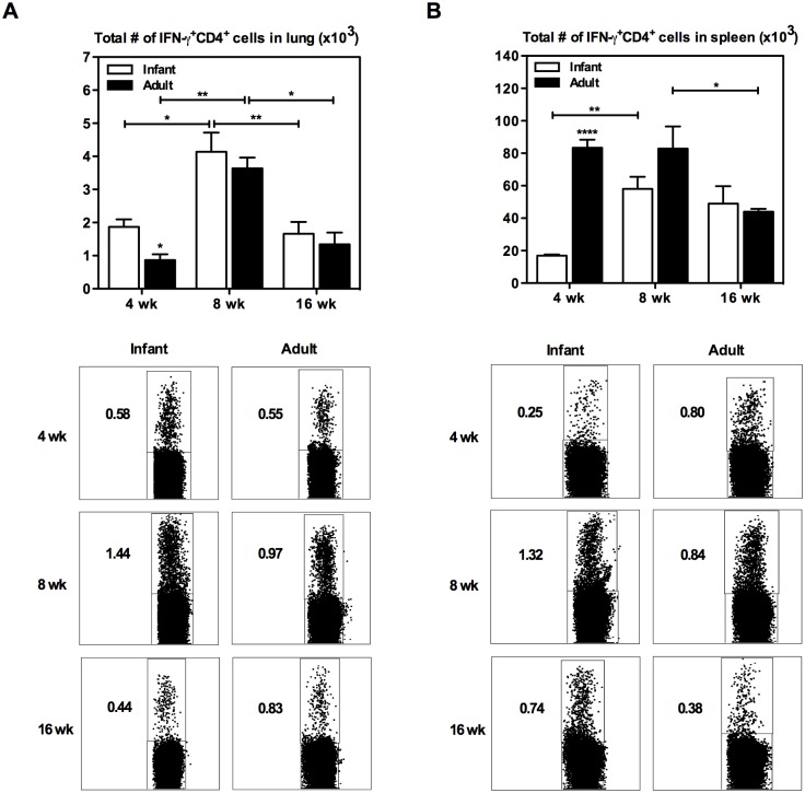
Fig 1. IFN-γ+CD4+ T cell kinetics in the lung and spleen of infant and adult mice following BCG immunization.
Infant and adult mice were immunized s.c. with BCG and sacrificed at 4, 8, or 16 weeks following immunization. Cells from the lung (A) and spleen (B) were isolated and stimulated with mixed M.tb culture filtrate (CF) and crude BCG (cBCG) for 24h or with media only as a control (unstimulated). Cells were stained with extracellular cell markers, followed by intracellular staining for IFN-γ, and analyzed by flow cytometry. Absolute numbers of IFN-γ+CD4+ T cells in the tissues were calculated (unstimulated numbers were subtracted from stimulated), and representative dot plots are shown. Results are from one to two independent experiments per timepoint, n = 4-8/group/timepoint. Data are expressed as Mean ± SEM. *, p < 0.05; **, p < 0.005; ****, p < 0.0001.

